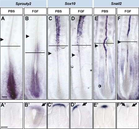
Figure 4.
Ectopic FGF inhibits NCC specification and emigration. (A–F) Stage HH11 and 12 embryos were exposed to heparin-coated beads either with PBS (A, C, and E) or with FGF4 (B, D, and F) after 16–18 h in culture. In situ hybridization analysis of Sprouty, Sox10, and Snail2 expression. Black arrowheads point to the last formed somite. (A’–F’) Transversal sections through the region where the bead was located, indicated by horizontal lines in the corresponding figures. Black arrows point to ectopic expression in B’ and to down-regulation of expression in D’ and F‘. Bars: (A, for A–F) 100 µm; (A’, for A’–F’) 40 µm.

