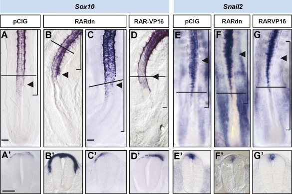
Figure 6.
RA signaling is required at the neural tube to control the timing of NCC emigration. Chicken embryos at stages HH11–13 electroporated on the right neural hemitube with control pCX-IRES-GFP construct (pCIG), a dominant-negative truncated version of RAR-α (pCIG-RARdn), or a constitutively active form of RAR-α (RAR-VP16) and analyzed 18–24 h later. In situ hybridization of Sox10 (A–D) and Snail2 expression (E–G). Black arrowheads point to the last formed somites, and brackets indicate the electroporated area. (A’–G’) Transversal sections at the level are indicated by horizontal lines in the corresponding figures. Bars: (A, for A, B, and D) 100 µm; (C, for C and E–G) 80 µm; (A’, for A’–G’) 45 µm.

