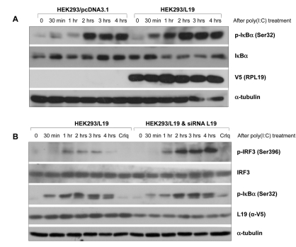
Figure 3.
Effect of L19 on activation of IRF3 or IκBα in TLR3 signaling. (A) HEK293 cells grown in 60mm plates were transfected with control (pcDNA3.1) or V5-tagged L19. 48 hrs after transfection, cells were stimulated with 20µg/ml poly(I:C) for the indicated time. Phosphorylated IκBα (p-IκBα) and total IκBα were determined by western blotting using anti-p-IκBα (Ser32) and anti-IκBα antibodies. The expression level of L19 in the cell lysates was analyzed with anti-V5 antibody. To confirm equal loading, membranes were re-probed with anti-α-tubulin antibody. (B) siRNA oligo targeting L19 or negative control were transfected into L19 expressing HEK293 (HEK293-L19) cells to silence the expression of L19. 48 hrs after transfection, cells were stimulated without or with 20µg/ml of poly (I:C) for the indicated time. Total cell lysates analyzed by immunoblotting with anti-phospho-IRF3 (Ser396) or anti-IRF-3 antibody, anti-p-IκBα (Ser32) or anti-IκBα antibodies. The efficiency of L19 RNAi was confirmed by immunoblotting with anti-V5 (L19) antibody.

