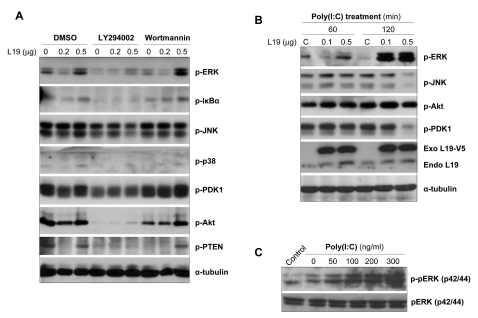
Figure 5.
Effects of PI3K inhibitors together with L19 on TLR-mediated activation of intracellular signaling molecules. (A) A172 cells were transfected with empty vectors or V5-tagged L19 plasmid. 18 hrs after transfection, cells were stimulated with poly(I:C) (25µg/ml) in the presence or absence LY294002 (LY, 20µM) and wortmannin (Wort, 5µM) for 20 hrs and then total cell lysates analyzed by immunoblotting with anti-phospho-ERK, -IκBα, -JNK, -p38, -PDK1, -Akt, -PTEN antibodies. To confirm equal loading, membranes were re-probed with anti-α-tubulin antibody. (B) A172 cells were transfected with empty vectors or V5-tagged L19 plasmid. 20 hrs after transfection, cells were stimulated with poly(I:C) (25µg/ml) for 60 or 120 min (B) and 12 hrs (C) and then total cell lysates analyzed by immunoblotting with anti-phospho-ERK, -JNK, -PDK1, -Akt antibodies. The expression level of exogenous or endogenous L19 in the cell lysates were analyzed with anti-L19 monoclonal antibody. To confirm equal loading, membranes were re-probed with anti-α-tubulin antibody.

