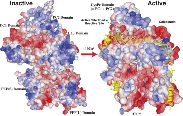
Figure 9.
Schematic of the 3D structure of inactive and active m-calpain. Surface-type schematic 3D structures of inactive (Ca2+ free) and active (Ca2+- and calpastatin-bound) forms of m-calpain using PDB data, 1KFX109) and 3DF0.111) The oligopeptides represented by the yellow ribbon + ball-and-stick indicate portions of calpastatin bound to active m-calpain. The dotted lines indicate portions that were too mobile for the 3D structure to be determined. The active protease domain (CysPc) is formed by the fusion of core domains PC1 and PC2 upon the binding of one Ca2+ to each of the core domains. The active site is circled in black. Blue balls represent Ca2+ (not all are visible).

