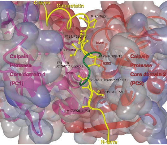
Figure 11.
Calpastatin region B binds and inhibits calpain. Enlarged view of the interaction between calpastatin (yellow) and the catalytic cleft in the protease core domain PC1 (pink)–PC2 (red) of m-calpain (the 3D structure is from 3DF0). Calpastatin binds in the substrate orientation indicated by positions P3 to P3′. At P1, calpastatin distorts from the substrate path and projects residues 174–178 or 613–617, which form a kink between the P2 and P1′ anchor sites.

