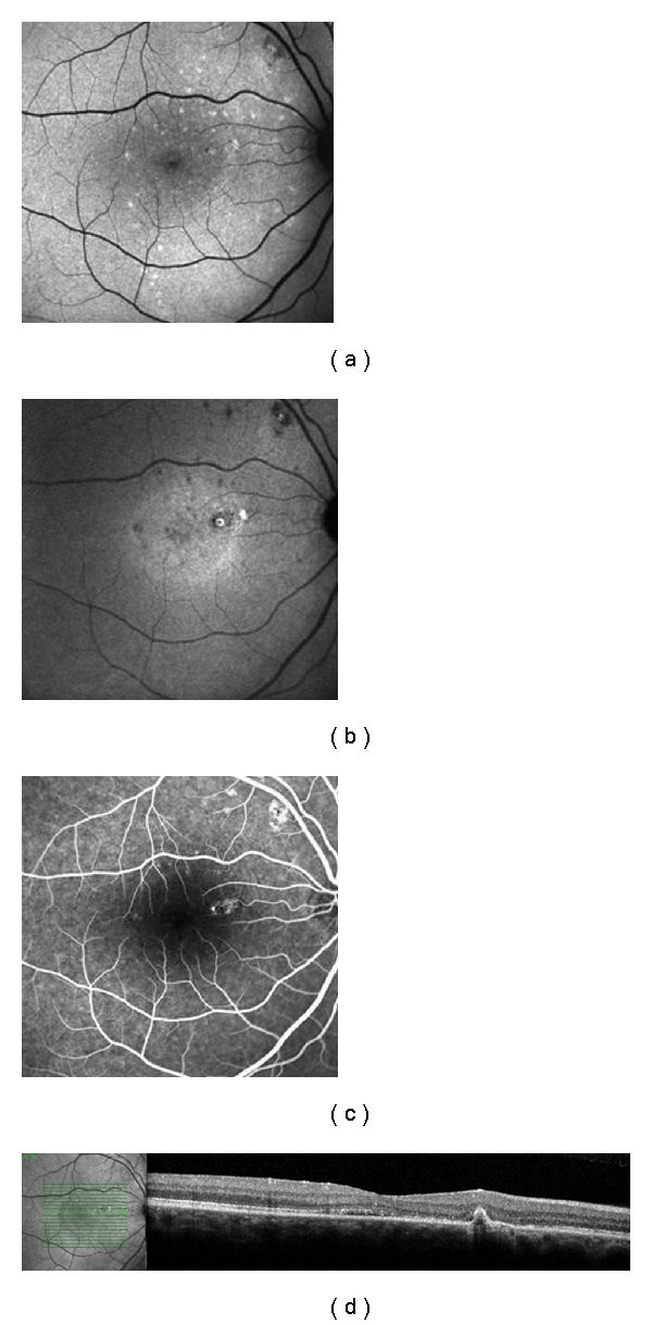
Figure 2.

Patient with 9 months of history of CSC in the right eye. (a) Standard FAF, showing mottled hyperFAF spots; (b) NIA, presenting macular and supratemporal to the optic disk hypofluorescent points—the macular spot surrounded by a ring of hyperAF, corresponding exactly with the PED in (d); (c) spots of leakage in the angiography; (d) SD-OCT exhibiting the PED.
