
Fig 2.
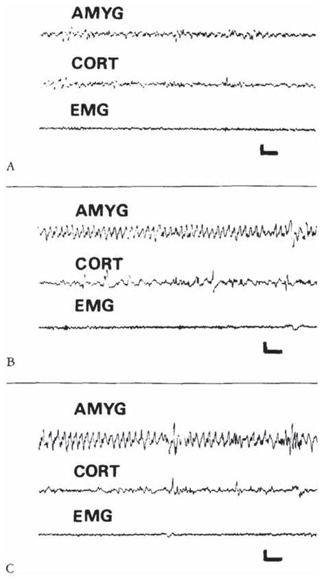
Electroencephalograms of a 9-day-old rat before corticotropin-releasing hormone (CRH) administration (A), and 9 and 120 minutes after intracerebroventricular infusion of 015 nm of CRH (B, C). The onset of semirhythmic slow wave discharges, confined to amygdala leads (AMYG). is evident in B. They persisted intermittently for several hours (C). CORT = cortical lead; EMG = motion-detecting electrodes placed over the angle of the jaw. Vertical bar = 50 μV; horizontal bar = 1 second.