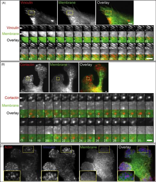
Figure 2.
A. Dynamics of the cell membrane at the focal adhesion of HFF transiently expressing GFP-Vinculin and membrane marker IL2R-mCherry. HFF were cultured on a 2D FN matrix. A representative focal adhesion is highlighted by the ROI. The ROI montage shows every 10th frame of the time lapse of focal adhesion extension; the total duration of time-lapse image acquisition used for the montage is 12.7 min. Scale bar indicates 3 μm. B. Dynamics of the cell membrane at a podosome of IC-21 macrophage transiently expressing GFP-Cortactin and membrane marker IL2R-mCherry. Transfected macrophages were cultured on a 2D FN matrix. The ROI montage shows every 10th frame, and the total duration of the time-lapse image acquisition used for the montage is 5.2 min. Scale bar indicates 1 μm. C. TIRF microscopy of IC-21 macrophage expressing IL2R-mCherry and immuno-labeled with phalloidin-Alexa488 and anti-vinculin Cy-5-conjugated antibody. Enlargements of the ROI are shown as insets.

