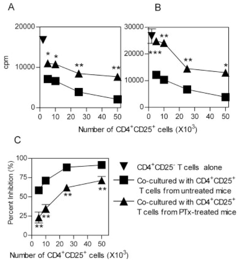
Figure 3.

In vivo PTx treatment reduces the suppressor effect of splenic CD4+CD25+ T cells. CD4+CD25+ and CD4+CD25− T cells were purified by flow cytometry, from MACS-purified CD4 cells derived from spleen. CD4+CD25− T cells (5 × 10⁴ cells/well) were mixed with increasing numbers of CD4+CD25+ T cells (5 × 10³–5 × 10⁴ cells/well). The cells were stimulated with APC (from normal control mice, 2 × 10⁵ cells/well) plus a soluble anti-CD3 antibody (0.5 μg/mL) and cultured for 72 h. Proliferation was measured by [3H]thymidine incorporation. The responder CD4+CD25− T cells were either from normal control mice (A) or from PTx-treated mice (B). The inverted triangle indicates CD4+CD25− T cells alone; the square indicates untreated mice derived-CD4+CD25+ T cells mixed with CD4+CD25− T cells; the triangle indicates CD4+CD25− T cells mixed with CD4+CD25+ T cells derived from PTx-treated mice. The data shown are representative of at least five separate experiments with similar results. (C) Percent inhibition of CD4+CD25+ T cells from normal control (squares) or PTx-treated (triangles) mouse spleen to the proliferation of normal control mouse CD4+CD25− T cells. The data shown are summarized from five separate experiments (with three to five mice per group). *p<0.05, **p<0.01, and ***p<0.001, compared with inhibition elicited by normal mouse CD4+CD25+ T cells.
