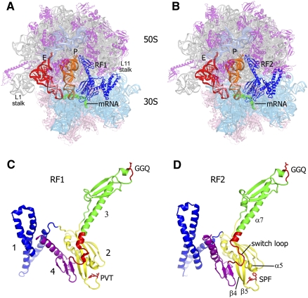
FIGURE 1.
Crystal structures of the 70S translation termination complexes bound with RF1 and RF2. (A,B) 3.2-Å and 3.0-Å structures of the 70S termination complexes bound with RF1 and RF2 (blue) in response to the UAA stop codon (mRNA is shown in green) and in the presence of deacylated P- (orange) and E-site (red) tRNAs. 23S rRNA is shown in gray, 5S rRNA in teal, 50S subunit proteins in magenta, 16S rRNA in cyan, and 30S subunit proteins in pink. (C,D) The structures of RF1 and RF2 in their ribosome-bound conformation, rotated ∼180° from A and B; the structures are colored according to their four-domain organization; GGQ, PVT, and SPF motifs and the switch loop are shown in red.

