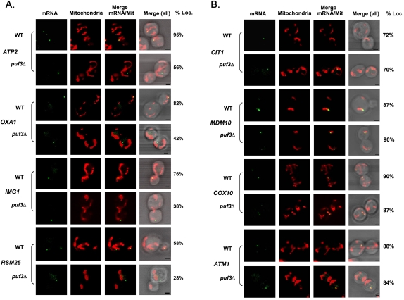
FIGURE 2.
Puf3-dependent and -independent mRNA localization to the mitochondria. (A) Puf3-dependent mRNA localization to the mitochondria. Representative confocal images of endogenously expressed MS2L-tagged mMPs in the puf3Δ background are shown. Cells cotransformed with plasmids expressing MS2-CP-GFP(x3) to visualize the mRNA and Oxa1-RFP to visualize mitochondria were grown to mid-log phase at 26°C on liquid synthetic medium containing glucose and then shifted to the same medium lacking methionine for 1 h. The percentage of RNA granules that colocalize with mitochondria (% Loc.) is given in %. (mRNA) Indicates the localization of GFP-labeled RNA granules. (Mitochondria) Indicates the localization of mitochondria labeled with Oxa1-RFP. (Merge mRNA/Mit) Indicates merger of the mitochondrial and mRNA windows. [Merge (all)] Indicates the merger between light, mitochondrial, and mRNA windows. Scale bar, 2 μm. (B) Puf3-independent mRNA localization to the mitochondria. Representative confocal images of endogenously expressed MS2L-tagged mMPs in the puf3Δ strain background are shown. Cells were grown to mid-log phase at 26°C on liquid synthetic medium and then shifted to medium lacking methionine for 1 h. Cells are labeled as described in A.

