
Figure 2.
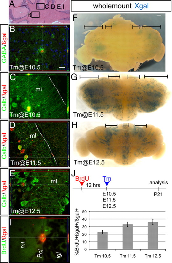
GABAergic cn interneurons and Purkinje cells are the first cell types marked by Ascl1CreER GIFM. A, Coronal section of the adult cerebellum highlighting where images in B–E and I were taken. B, Double-labeling immunohistochemistry for GABA (green) and βgal (red) shows that cn interneurons were marked when Tm was administered at E10.5. Anti-calbindin (green) and anti-βgal (red) double-labeling immunohistochemistry shows that fate-mapped cells are Purkinje cells when Tm was administered at E10.5 (C), E11.5 (D), and E12.5 (E). F–H, Dorsal views of whole-mount Xgal staining of P21 cerebella when Tm was administered at E10.5 (F), E11.5 (G), and E12.5 (H). I, BrdU was administered 12 h before Tm administration at E10.5, E11.5, or E12.5 and analysis performed at P21. Double-labeling immunohistochemistry for βgal (red) and BrdU (green) shows that Ascl1CreER GIFM-marked Pcs retained BrdU labeling. J, Quantification of the number of double-positive BrdU and βgal Pcs (mean ± SEM; n = 3; unpaired t test). Pcl, Purkinje cell layer. Scale bar: A, 100 μm; B–E, I, 20 μm; F–H, 600 μm.