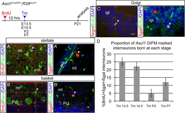
Figure 5.
A population of cerebellar interneurons is born during embryonic development. BrdU was administered 12 h before Tm administration at E13.5, E14.5, P2, and P7. A, Double-labeling immunohistochemistry in the outer ml for BrdU (red) and parvalbumin (green) shows some stellate cells are born at E14.5. A′, Double-labeling immunohistochemistry for βgal (red) and BrdU (green) shows an Ascl1CreER GIFM fate-mapped stellate cell born embryonically. B, Double-labeling immunohistochemistry in the inner ml for BrdU (red) and parvalbumin (green) shows some basket cells are born at E14.5. B′, Double-labeling immunohistochemistry for βgal (red) and BrdU (green) shows an Ascl1CreER GIFM fate-mapped basket cell born embryonically. C, Double-labeling immunohistochemistry in the igl for BrdU (green) and neurogranin (red) shows some Golgi cells are born at E14.5. C′, Double-labeling immunohistochemistry for βgal (red) and BrdU (green) shows an Ascl1CreER GIFM fate-mapped Golgi cell born embryonically. D, Quantification of the number of BrdU and βgal double-positive interneurons marked at each time point with Ascl1CreER GIFM is shown (mean ± SEM; n = 3; unpaired t test). Pcl, Purkinje cell layer; pia, pial surface. Scale bar, 20 μm.

