Figure 7.
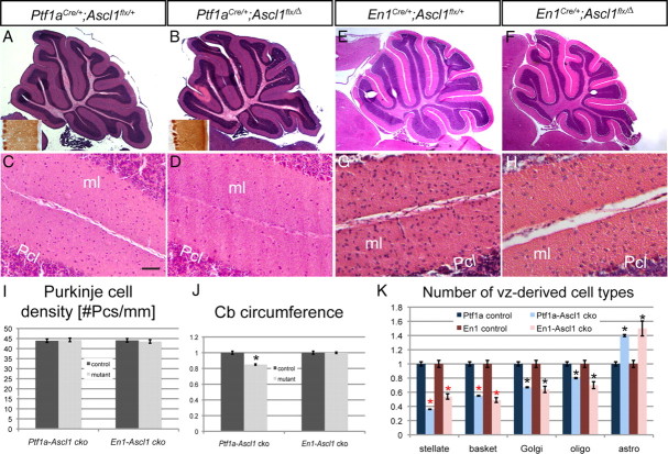
Conditional deletion of Ascl1 in the cerebellum differentially affects the number of interneurons and glia in the adult. Hematoxylin and eosin (H&E)-stained sections show smaller cerebellum in a Ptf1a-Ascl1 cko (B) compared with a littermate control (A). The insets in A and B are immunostaining for calbindin showing no difference in the overall morphology of Pcs. H&E-stained sections of En1-Ascl1 ckos (F) show a similar cellular phenotype to Ptf1a-Ascl1 ckos (A). Higher magnification images show severe reductions in the number of ml interneurons in both ckos (D, H) compared with littermate controls (C, G). I, Quantification of the number of Purkinje cells per millimeter in Ptf1a-Ascl1 and En1-Ascl1 ckos and littermate controls. J, Quantification of the average cerebellar circumference in Ptf1a-Ascl1 and En1-Ascl1 ckos and littermate controls with controls set as 1 shows significantly smaller cerebellum size in Ptf1a-Ascl1 ckos (p = 0.01) but not En1-Ascl1 ckos. K, Quantification of the GABAergic interneurons and glia in Ptf1a-Ascl1 and En1-Ascl1 ckos and littermate controls with average control numbers set as 1 (mean ± SEM; Ptf1a-Ascl1 cko, n = 3; Ptf1a-Ascl1 control, n = 3; En1-Ascl1 cko, n = 3; En1-Ascl1 control, n = 3; unpaired t test; the red asterisk depicts p < 0.003; the black asterisk, p < 0.05). Scale bar: A, B, E, F, 100 μm; C, D, G, H, 20 μm.

