Fig. 3.
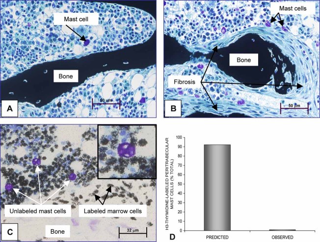
(A) cPTH increases mast cell number at the bone–bone marrow interface in 6-month-old female rats without increasing mast cell proliferation. Peritrabecular fibroblasts were not present, and mast cells were located predominantly in bone marrow in the control rats. (B) On day 7 of treatment with cPTH, most cancellous bone surfaces were covered by multiple layers of fibroblasts, and mast cells commonly were located adjacent to the fibroblasts. Specimens shown in panels A and B are undemineralized, embedded in plastic, and stained according to von Kossa with tetrachrome counterstain. (C) Although continuous [3H]thymidine labeled many bone marrow cells in 6-month-old female rats, very few mast cells were labeled (JB-4-embedded, toluidine blue–stained section). A higher-magnification insert clearly shows the absence of labeling in a representative mast cell that is surrounded by highly labeled and unlabeled marrow cells. (D) The predicted number of labeled mast cells assuming proliferation is required for mast cell migration to bone surfaces. The observed number was much lower, indicating that treatment with cPTH results in the migration of postproliferative mast cells to bone surfaces. The predicted value was determined as described previously.(17)

