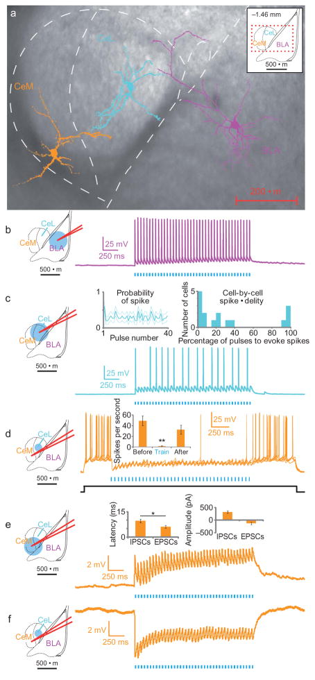
Figure 2. Projection-specific excitation of BLA terminals in the CeA activates CeL neurons and elicits feed-forward inhibition of CeM neurons.
a) Two-photon images of representative BLA, CeL and CeM cells imaged from the same slice, overlaid on a brightfield image. (b–f) Schematics of the recording and illumination sites for the associated representative current-clamp traces (Vm=~−70 mV. b) Representative BLA pyramidal neuron trace expressing ChR2, all of which spiked for every pulse (n=4). c) Representative trace from a CeL neuron in the terminal field of BLA projection neurons, showing both sub- and supra-threshold excitatory responses upon photostimulation (n=16). Inset left, population summary of mean probability of spiking for each pulse in a 40-pulse train at 20Hz, dotted lines indicate s.e.m. Inset right, frequency histogram showing individual cell spiking fidelity; y-axis is the number of cells per each 5% bin. d) Six sweeps from a CeM neuron spiking in response to a current step (~60 pA; indicated in black) and inhibition of spiking upon 20Hz illumination of BLA terminals in the CeL. Inset, spike frequency was significantly reduced during light stimulation of CeL neurons (n=4; spikes/second before (49±9.0), during (1.5±0.87), and after (33±8.4) illumination; mean±s.e.m.). (e–f) Upon broad illumination of the CeM, voltage-clamp summaries show that the latency of excitatory postsynaptic currents (EPSCs) is significantly shorter than the latency of inhibitory postsynaptic currents (IPSCs), whereas there was a non-significant difference in the amplitude of EPSCs and IPSCs (n=11; *p=0.04, see insets). The same CeM neurons (n=7) showed either net excitation when receiving illumination of the CeM (e) or net inhibition upon selective illumination of the CeL (f).

