Figure 4.
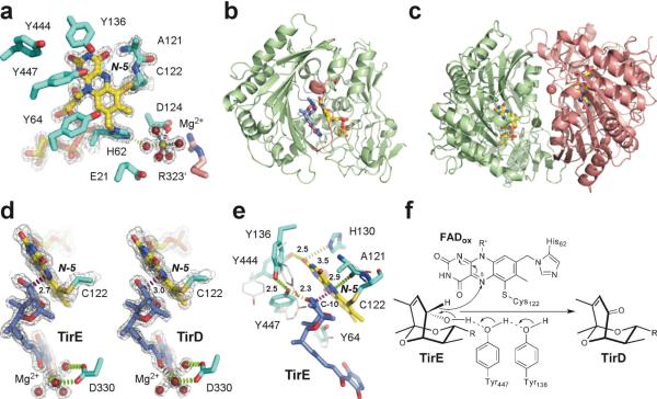
Ligand-free and substrate/product-bound TamL. (a) Catalytic and the Mg2+-binding sites in the substrate-free TamL with covalently bound FAD (yellow sticks) (PDB ID 2Y08). Residues from the same monomer are in cyan, from the symmetry-related monomer in pink. (b) Tirandamycin (blue sticks) in the active site of TamL (PDB ID 2Y3R). Residues 323–336 at the mouth of the substrate binding cleft are in pink. (c) Ribbon representation of TamL dimer formed by the green and pink monomers related by the non-crystallographic symmetry (PDB ID 2Y08). The Mg2+ atoms (spheres of matching colors) stabilize dimerization interface. (d) Interactions between the C-10 site of oxidation in TirE and the N-5 locus in FAD are highlighted in magenta dash line defining an angle with the N-5/N-10 flavin atoms of 110°. Installation of the keto group in TirD flattens the ketal ring pulling C-10 away from the N-5 atom by 0.3 Å (PDB ID 2Y3R). (e) Superimposition of the amino acid residues in the tirandamycin-bound (cyan sticks) compared to substrate-free (grey sticks) TamL. (f) Mechanism of dehydrogenation at C-10 in TirE. In all panels, oxygen atoms are in red, nitrogen in blue, sulfur in dark yellow, magnesium in green. Electron density 2Fo-Fc map (gray mesh) is contoured at 1.5 σ. Distances are in Angstroms.

