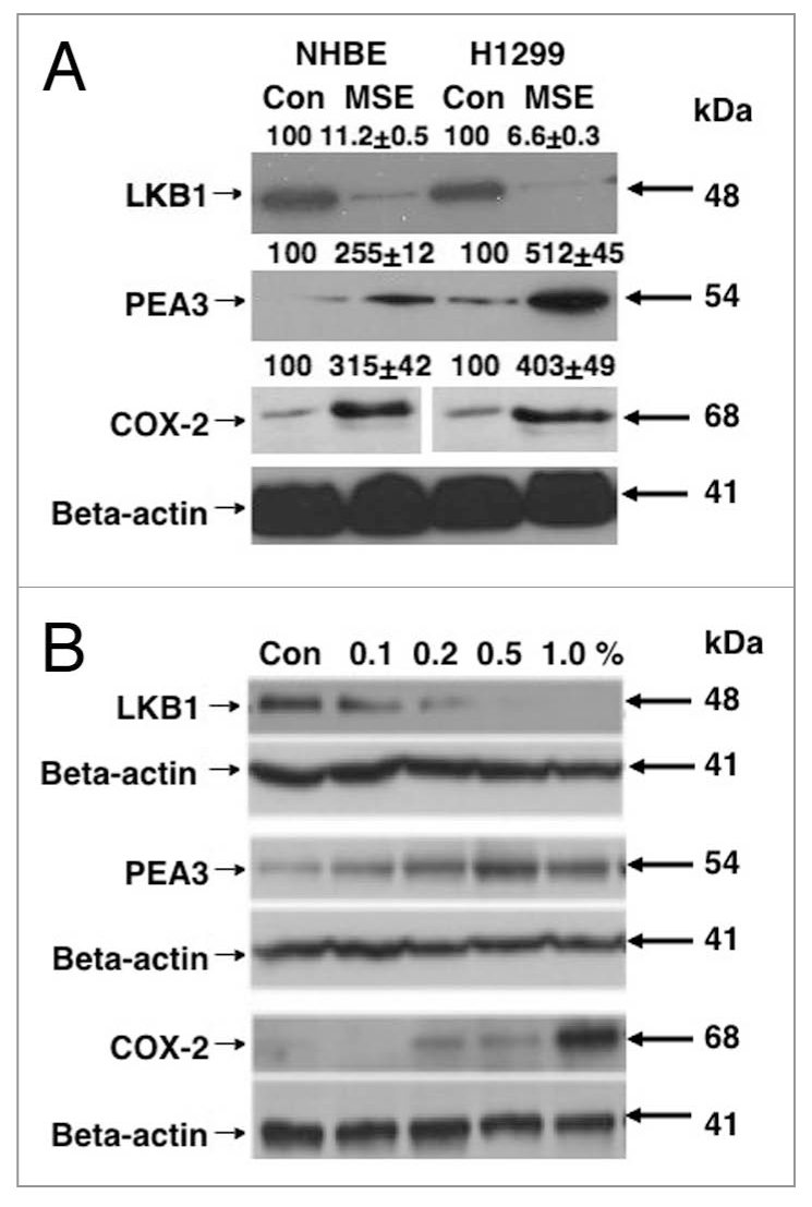
Figure 1.

MSE affects the levels for LKB1, PEA 3 and PTGS-2/COX-2 in normal lung and lung cancer cells. (A) NHBE normal lung and H1299 lung cancer cells were exposed to 0.5% MSE for 48 h. (B) H1299 lung cancer cells were incubated with MSE in a dose-dependent manner (Con, control, 0.2, 0.5, 1.0 and 2% MSE) for 24 h. Total lysates were analyzed by immunoblotting with antibodies against LKB1, PEA 3, PTGS-2/COX-2 and b-actin. Immunoblots were scanned and quantified by Image Quant software version 3.3 and normalized for the b-actin protein levels. Median data expressed as percentages of data obtained from control sample representing three independent experiments were shown above blot images. Statistical analysis was performed using a student t-test [16].
