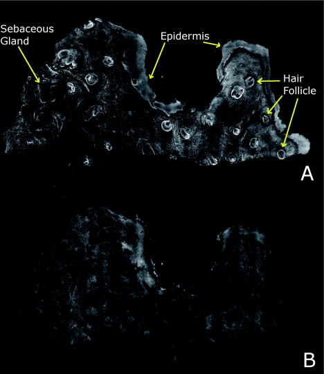
Figure 6.
Another example of a confocal mosaic of a Mohs excision of normal skin in (a) fluorescence mode showing the epidermis, sebaceous glands, eccrine glands, adipose tissue, and hair follicles. The tissue was stained with 0.6 mM acridine orange for 20 s. The corresponding mosaic in (b) reflectance mode highlights cellular cytoplasm and dermal collagen. The corresponding digitally stained confocal mosaic and histology is shown in Fig. 7.

