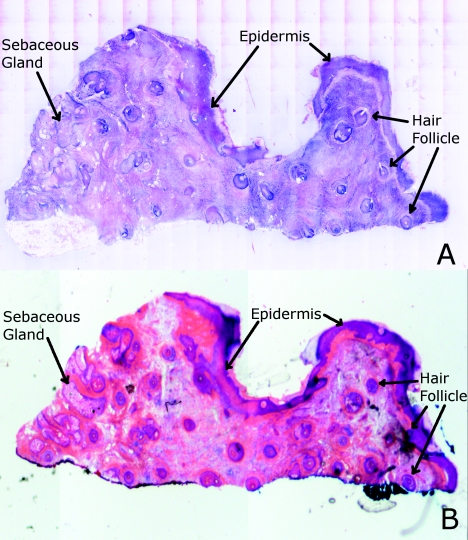
Figure 7.
Digitally stained confocal mosaic and histology for the excision of normal skin that was shown in Fig. 6. (a) Digitally stained mosaic shows the nuclear detail in purple and the cellular cytoplasm and dermis in pink, which is similar to (b) the corresponding histology.

