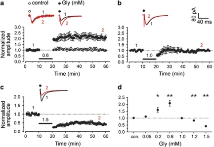
Figure 1.
Glycine induced bidirectional persistent changes in EPSCs in a dose-dependent manner. (a) Application of 0.6 mM glycine for 10 min in Mg2+-free ACSF caused significant potentiation of evoked AMPAR-mediated EPSCs when cells were held at −65 mV in a conventional whole-cell patch configuration (n=6). EPSCs were recorded in an ACSF perfusion medium containing bicuculline methiodide (BMI, 10 μM) to block GABAA receptor-mediated inhibitory synaptic currents. However, 10-min treatment of Mg2+-free ACSF alone failed to produce any persistent change in EPSCs (control). In this and all of the following figures, unless stated, ACSF was always kept in 1.0 mM Mg2+ before and after glycine treatment, and traces above the graph show averaged AMPAR-mediated EPSCs chosen at the times indicated on the graph. (b) When the concentration of glycine increased to 1.0 mM, no persistent change in EPSCs was observed (n=6). (c) In all, 1.5 mM glycine treatment in Mg2+-free ACSF induced LTD of AMPAR-mediated EPSCs (n=6). (d) Summary of data comparing persistent changes in EPSCs induced by glycine across a range of concentrations (0.05, 0.2, 0.6, 1.0, 1.2, and 1.5 mM). *P<0.05; **P<0.01, compared with control, ANOVA LSD test.

