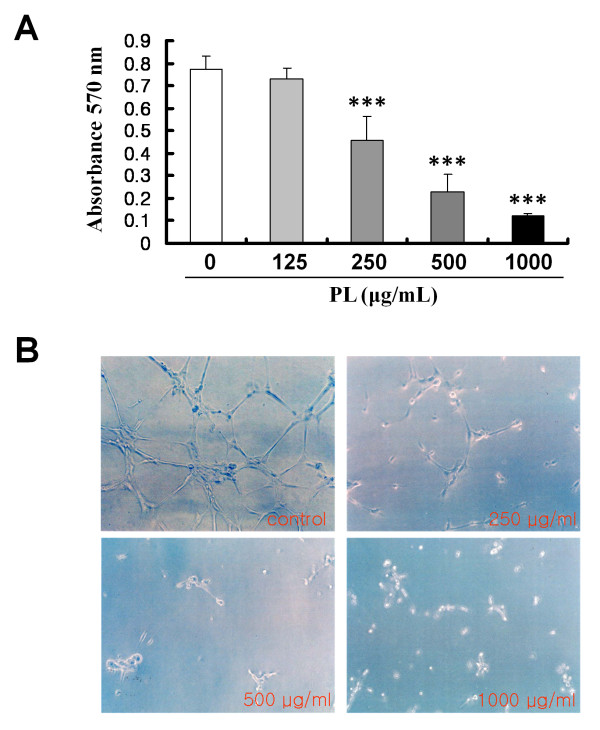
Figure 3.
Effect of PL on proliferation of HUVECs and capillary tube formation. A, HUVECs were seeded in 0.2% gelatin-coated wells of 96-well plates at an initial density of 5 × 10³ cells/well. On the next day, the indicated concentrations of PL were added to each well for 24 h, and cell numbers were determined using the MTT assay as described in Methods. The data shown represent the mean ± SD of 3 independent experiments (***, P < 0.001). B, PL inhibited capillary tube formation by HUVECs on Matrigel. HUVECs were seeded at a density of 104 cells/well in 1:2-diluted Matrigel-coated 24-well plates and incubated with the indicated concentrations of PL. At 18 h, the control HUVECs had formed an interconnected network of anastomosing cells (which had a honeycomb appearance), whereas HUVECs treated with PL showed a significant reduction in tube formation in a dose-dependent manner. Photomicrographs were taken using an inverted phase-contrast microscope.

