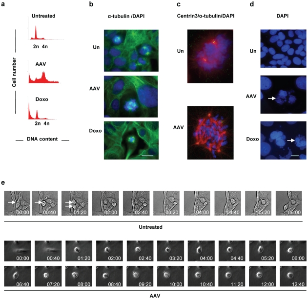
Figure 1. U2OSp53DD cells infected with AAV die by mitotic catastrophe.
(a) U2OSp53DD cells die 4 days after infection. Cells were infected with AAV and analyzed by PI staining and FACS 4 days post-infection (x-axis: DNA content; y-axis: cell count). Doxorubicin (Doxo) leads to cell death in mitosis and was used as a control. (b) AAV-infected cells show multiple spindle poles. U2OSp53DD cells were infected and then stained for α-tubulin 4 days post-infection. Bar: 30 µm. (c) Infected and control uninfected cells were stained for DNA with DAPI, for α-tubulin (red) and for centrin3 (green). Merged red and green give yellow. (d) U2OSp53DD cells were infected with AAV and then stained with DAPI 4 days post-infection. The arrows highlight micronucleated cells. Bar: 15 µm. (e) Prolonged mitosis can lead to cell death. Cells were infected with AAV and analyzed by time-lapse brightfield-microscopy 2 days post-infection. Images were acquired using the 20× objective and phase-contrast was used for the infected cells. The arrows indicate a normally-dividing cell.

