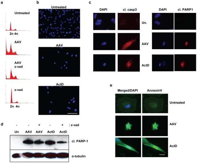
Figure 6. AAV induces apoptosis in M059K cells.
(a) Inhibition of caspases leads to a decrease in the SubG1 population induced by AAV. M059K cells were infected, treated with zVAD-fmk and then stained with PI and analyzed by flow cytometry 4 days after infection (x-axis: DNA content; y-axis: cell count). Treatment with zVAD-fmk alone did not have a significant effect on the cells. (b) Infection with AAV does not lead to micronucleation in M059K cells. Infected cells were stained with DAPI 4 days post-infection. ActD was used as a control with no micronucleation and positive for apoptosis. Bar: 115 µm. (c) AAV-infected M059K cells have condensed or fragmented chromatin and are positive for cleaved caspase-3 and cleaved PARP-1. Cells were infected and analyzed by IF 4 days post-infection. DAPI was used to stain the nuclei. Bar: 35 µm. (d) Western analysis showing that glioblastoma cells are positive for cleaved PARP-1 after infection with AAV. Protein levels were assayed 4 days after infection. α-tubulin was used as a loading control. (e) AAV-infected M059K cells are positive for annexin-V staining. Cells were analyzed by IF 4 days post-infection, as in Figure 4. DNA was stained with DAPI. Bar: 30 µm.

