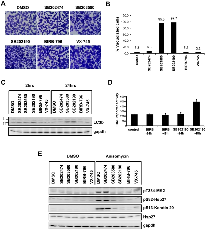
Figure 5. p38 MAPK inhibition is neither sufficient for induction of autophagy nor for FHRE reporter activation.
(A) HT29 cells were treated for 24 hours with SB203580 (5 µM), SB202190 (5 µM), SB202474 (5 µM), BIRB-796 (1 µM), VX- 745 (2 µM) and DMSO control. Cells were fixed in 95% ethanol and stained with crystal violet (original magnification 600×). (B) The percentage of vacuolated cells from three independent images was calculated and the mean values are shown. (C) Cells treated with the set of inhibitors as indicated were analyzed by Western blotting for the autophagic marker – LC3b. LC3-I and its lipid conjugated form- LC3-II are indicated. Gapdh was used as loading control. (D) HT29 cells stably transfected with FHRE-luc reporter were treated with 10 µM SB202190 or 1 µM BIRB-796 for indicated time points and luciferase activity measured and normalized to protein content. (E) Cells were treated with the respective inhibitors or DMSO for 1 hour as indicated, followed by 30 minutes treatment with anisomycin (10 µg/ml) or DMSO. For confirming p38 inhibition, the phosphorylations of MK2, keratin 20 and Hsp27 were monitored by Western blotting. Total Hsp27 and gapdh were used as loading controls.

