
Fig. 5.
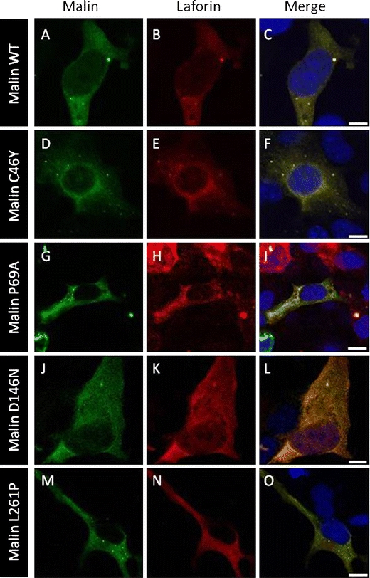
Malin mutants colocalize with laforin. COS-7 cells were transiently cotransfected with the GFP-malin and laforin-Myc plasmids. Direct visualization of GFP-malin (green) and immunofluorescence with anti-Myc (red) was performed. Most representative localizations with wild-type (WT) malin (a–c), C46Y (d–f), P69A (g–i), D146N (j–l), and L261P (m–o) mutants are shown. Magnification bars, 10 μm