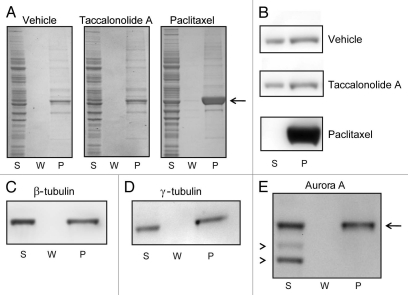
Figure 3.
Effects of paclitaxel or taccalonolide A on tubulin polymer formation in cytosolic extracts. HeLa cell lysates were collected, chilled to depolymerize pre-existing microtubules and then incubated with vehicle, 20 µM paclitaxel or 20 µM taccalonolide A for 30 min at 37°C. Microtubule polymer was separated from soluble tubulin by centrifugation at room temperature. The total protein (A) and β-tubulin (B) levels present in the supernatant (S), wash (W) and pellet (P) were determined by total protein staining or β-tubulin immunoblotting, respectively. The location of tubulin in the total protein stained gel is indicated with an arrow (3A). β-tubulin (C) and the microtubule associated proteins γ-tubulin (D) and Aurora A (E, arrow), were detected in the microtubule containing pellet (P) of samples treated with 100 µM taccalonolide A as compared to non-specific background bands, which were retained in the supernatant (E, arrowheads).

