
Figure 1.
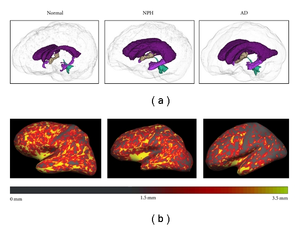
(a) Three-dimensional representation of the ventricles in normal, NPH, and AD participant (left to right). The ventricles of the NPH and the AD participants are enlarged relative to the normal participant. (b) FreeSurfer's cortical thickness maps in the same normal, NPH, and AD participant (left to right) are shown. The cortex of the AD participant is notably thinner, particularly in posterior regions, than that of the normal and NPH participant.