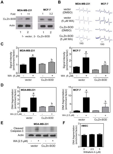
Figure 2. Cu,Zn-Superoxide dismutase (Cu,Zn-SOD) overexpression attenuates withaferin A (WA)-induced apoptosis in MDA-MB-231 and MCF-7 cells.
(A) Immunoblotting for Cu,Zn-SOD using lysates from MDA-MB-231 or MCF-7 cells stably transfected with empty vector (lane 1) or vector encoding for Cu,Zn-SOD (lane 2). (B) Representative EPR spectra in MDA-MB-231 and MCF-7 cells stably transfected with empty vector or vector encoding for Cu,Zn-SOD and treated for 4 h with DMSO or 5 µM WA. (C) Quantitation of EPR signal intensity in MDA-MB-231 and MCF-7 cells transfected with empty vector or vector encoding for Cu,Zn-SOD and treated for 4 h with DMSO or 5 µM WA. Results shown are mean ± SD (n = 3). (D) Histone-associated DNA fragment release into the cytosol (a measure of apoptosis) in MDA-MB-231 and MCF-7 cells transfected with empty vector or vector encoding for Cu,Zn-SOD and treated for 24 h with DMSO or WA. Results shown are mean ± SD (n = 3). (E) Immunoblotting for cleaved caspase-3 using lysates from MDA-MB-231 cells stably transfected with empty vector or vector encoding for Cu,Zn-SOD and treated for 24 h with DMSO or WA. (F) Histone-associated DNA fragment release into the cytosol in HMEC treated for 24 h with DMSO or WA. Results shown are mean ± SD (n = 3). Significantly different (P<0.05) compared with acontrol (DMSO-treated), and b between groups at each dose by one-way ANOVA followed by Bonferroni's adjustment. For data in panels C,D, and F, data are shown as enrichment factor relative to DMSO-treated control. All experiments were repeated at least twice.

