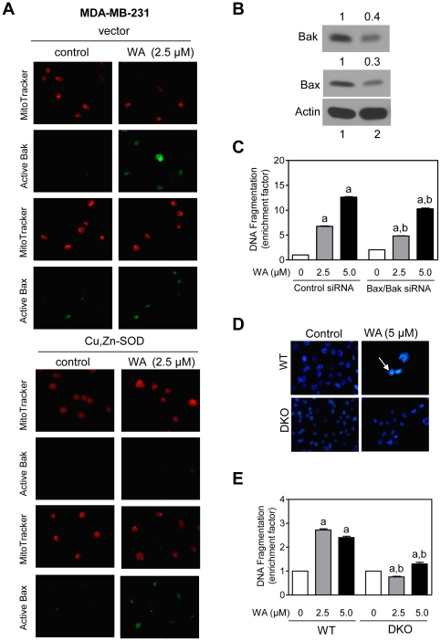
Figure 8. Bak and Bax are required for withaferin A (WA)-induced apoptosis.
(A) Immunofluorescence microscopy for active Bak and Bax in MDA-MB-231 cells stably transfected with empty vector or vector encoding for Cu,Zn-SOD and treated for 24 h with DMSO or WA. (B) Immunoblotting for Bak and Bax using lysates from MCF-7 cells transiently transfected with a control nonspecific small interfering RNA (siRNA; lane 1) or Bax- or Bak-targeted siRNA (lane 2). (C) Histone-associated DNA fragment release into the cytosol in siRNA-transfected MCF-7 cells following 24 h treatment with DMSO (control) or the indicated concentrations of WA. Results are shown as enrichment factor relative to DMSO-treated control siRNA transfected cells (mean ± SD, n = 3). (D) Fluorescence microscopic analysis for apoptotic cells with condensed and fragmented DNA (DAPI assay) in SV40 immortalized mouse embryonic fibroblasts (MEF) derived from wild-type (WT) and Bax and Bak double knockout (DKO) mice and treated for 24 h with DMSO (control) or 5 µM WA. (E) Histone-associated DNA fragment release into the cytosol in WT and DKO treated for 24 h with DMSO (control) or the indicated concentrations of WA. Results are shown as enrichment factor relative to DMSO-treated wild-type MEF (mean ± SD, n = 3). Significantly different (P<0.05) compared with aDMSO-treated control siRNA-transfected MCF-7 cells (panel C) or DMSO-treated WT MEF (panel E), and bbetween groups at each dose by one-way ANOVA followed by Bonferroni's test. Similar results were observed in two independent experiments.

