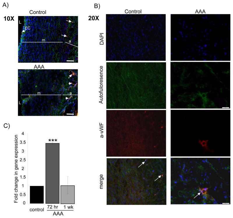
Figure 5.
Identification of capillaries using immunostaining with Von-Willebrand factor antibody (red), nuclei counterstained with DAPI (blue), elastin auto fluorescence (green). A) Endothelial cells identified lining the non-injured aortic endothelium in the suprarenal control segment (arrow), these are absent in the injured (AAA) aortic segment. Intact elastin shown in suprarenal control compared to disarranged architecture of the aortic wall in the injured segment with fraying of elastin fibers. L=lumen, EC= endothelial cells, m=media and a=adventitia. Scale bar = 100 μmB) Enhanced capillary density was observed in the AAA segment, representative image with arrow pointing to capillary in AAA aortic tissue compared to control. Scale bar = 50 μm. C) qRT-PCR: Fold change in gene expression of VEGFA in AAA tissue compared to non-injured control at 72 hours and one week after MSC injection. * Indicates a p-value <.05.

