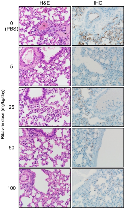
Figure 3. Histological analysis of lungs from Andes virus infected ribavirin treated and control hamsters.
Hamsters were infected with a lethal dose of Andes virus and treated daily with ribavirin at the indicated concentrations from day 1 through day 10 post-infection. Shown are hematoxylin and eosin (H&E) and immunohistochemistry (IHC) stained sections of lungs collected at day 8 post-infection from treated and control hamsters. Histological abnormalities were only noted in control hamsters which demonstrated perivascular edema (see *). IHC with a monoclonal antibody (shown) and polyclonal sera (not shown) revealed drastic reductions in the detection of Andes virus nucleoprotein in all ribavirin treated hamsters compared with the diffuse staining noted in the lung endothelium of control (PBS) treated animals.

