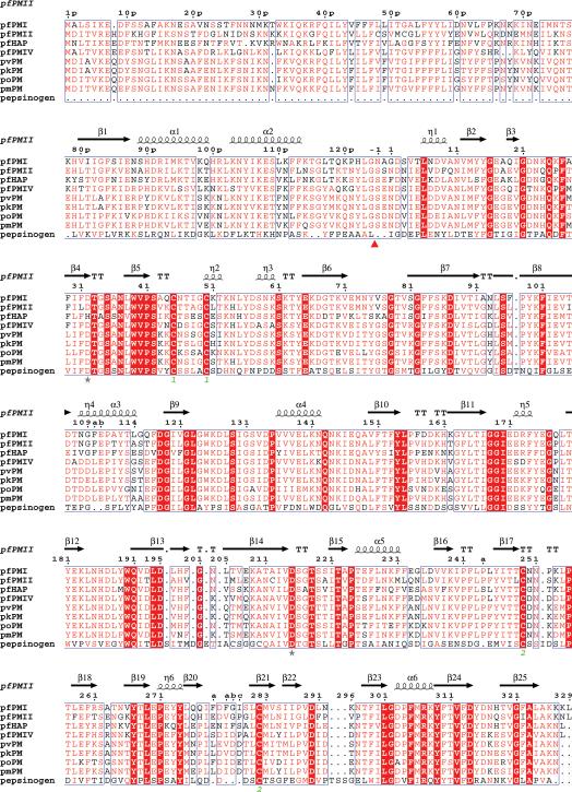
Figure 1.
Structure-based sequence alignment of proplasmepsins from different human malaria parasites with porcine pepsinogen. The Plasmodium species in this alignment are P. falciparum (pf), P. vivax (pv), P. ovale (po), P. malariae (pm), and P. knowlesi (pk). The alignment was performed with ClustalW [83] and the secondary structure was plotted using ESPript [84]. Similar residues identified by ESPript (global score=0.5) are shown in red letters and identical residues are highlighted by red background. The secondary structural elements as seen in the structure of the zymogen form of pfPMII (1PFZ) are drawn above the sequences. The catalytic residues are marked by stars. The disulfide links are identified as green numbers below the corresponding cysteine residues. The in vivo cleavage site of the prosegment is shown by a red triangle.

