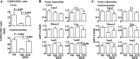
Fig. 4.
Deficiency of insulin secretion caused by STZ prevents the HFD-induced oxidative stress in the liver and skeletal muscle. A: the ratio of reduced (GSH) and oxidized glutathione (GSSG) was evaluated in the liver and gastrocnemius of the mice described in Fig. 1. Expression of genes involved in oxidative stress was determined by real-time PCR in the liver (B) and gastrocnemius (C) of the mice described in Fig. 1 and normalized to 36B4. Results represent means ± SE. Veh: saline. *P < 0.05, Veh + HFD vs. ND. #P < 0.05 and ##P < 0.01, STZ + HFD vs. Veh + HFD. Gpx1, glutiathone peroxidase 1; Cs, catalase; Gsr, glutiathone reductase; Hmox1, heme-oxygenase-1; Sod1, and -2 superoxide dismutase-1 and -2.

