Fig. 1.
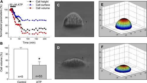
ATP causes persistent shrinkage of C11-Madin-Darby canine kidney (MDCK) cells. A: representative traces showing the time course of changes in height, surface area, and volume in cell superfused with 50 μM ATP for 30 min under isosmotic conditions. Initial height, surface, and volume values were taken as 100%. B: quantitative cell-volume changes in cells subjected to 60-min superfusion with isosmotic medium (control), or for 30 min with isosmotic medium containing 50 μM ATP, followed by 30 min with ATP-free medium (ATP). Initial cell volume values were measured before the addition of ATP or vehicle and varied in the 4,268 ± 246 μm3 range (n = 53). In all cases, individual initial cell volumes were taken as 100%. *P < 0.05 vs. control. C and D: light-microscope side-view images of the same cell immediately before ATP addition (C) and after 30-min treatment with 50 μM ATP, followed by 30-min incubation in ATP-free medium (D). E and F: perspective view of 3D models corresponding to the images in C and D, which were produced by the dual-image surface reconstruction (DISUR) technique, as described in materials and methods.

