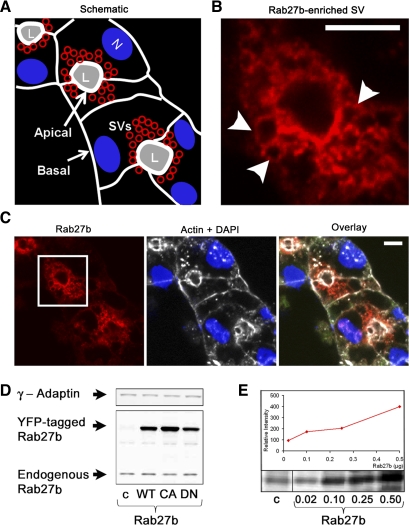
Fig. 2.
Rab27b expression is enriched on membranous structures in the subapical region of LG acinar cells. A: schematic of the reconstituted LG acinar cluster (in C) shows polarized cells that are delineated by a white outline representing cortical actin-enriched cell boundaries. The APM, which has a more highly enriched subapical actin network than the basolateral membrane, borders the lumen (L). SV (red) are located in the subapical regions, whereas nuclei (N) are more basal. B: high-magnification image of Rab27b immunofluorescence from C shows expression on apparent SV membranes in the subapical region. Arrows point to punctate structures. Bar = 5 μm. C: indirect immunofluorescence of reconstituted LG acinar cells probing for Rab27b (red), with a boxed region representing the image magnified in B. *, lumen. Endogenous Rab27b is highly enriched in the subapical region. Actin (white) and the nuclei (DAPI; blue) are shown for orientation. *, lumen; bar = 5 μm. D: fresh mouse LG lysate was prepared in RIPA buffer with protease inhibitors. Lysate protein (10 μg) from non-transduced (c) or mutant Rab27b-transduced (WT, CA, DN) LG acinar cells were resolved by SDS-PAGE and analyzed by Western blotting. Endogenous Rab27b is detected at its predicted molecular mass (∼29 kDa), and in transduced cells the tagged protein is detected at 52 kDa. γ-Adaptin was used to control for equal loading. E: serial dilution of purified recombinant His-tagged Rab27b protein in micrograms compared with endogenous Rab27b in 20 μg of lysate. Exposure to secondary antibody alone showed no bands in this region (data not shown).

