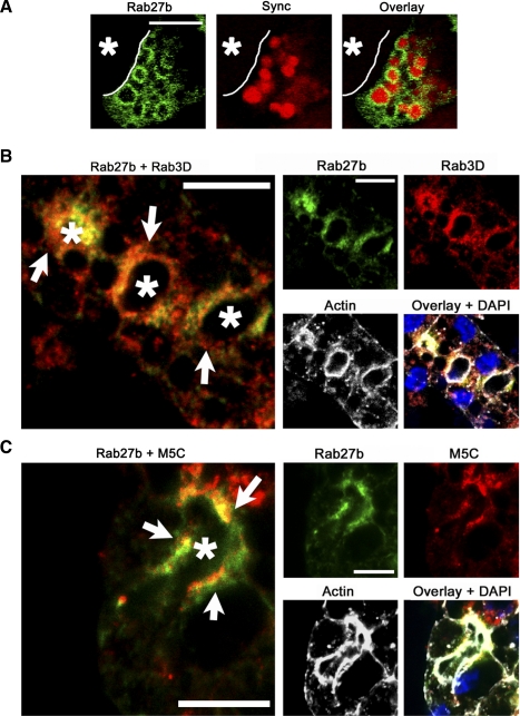
Fig. 3.
Rab27b colocalization with SV markers is consistent with a role in LG acinar exocytotic processes. A: reconstituted LG acinar cells co-transduced with Ad constructs encoding YFP-tagged WT (green) (MOI 3–5) and a GFP-tagged syncollin construct (red) (MOI 5) show that, in cells expressing both proteins, Rab27b is enriched on membranes of apparent SV containing syncollin-GFP. Bar = 5 μm. B: immunofluorescence detection of endogenous Rab27b (green) and Rab3D (red) reveal high co-localization (arrows), although Rab3D appeared to label a larger pool of vesicles. C: immunofluorescence detection of endogenous Rab27b (green) and myosin5C (red) in LG acinar cells also showed a high degree of colocalization (arrows). Nuclei are shown in the overlay images and were detected with DAPI (blue). Actin is shown in white. Bar = 10 μm. *, Lumen.

