Fig. 4.
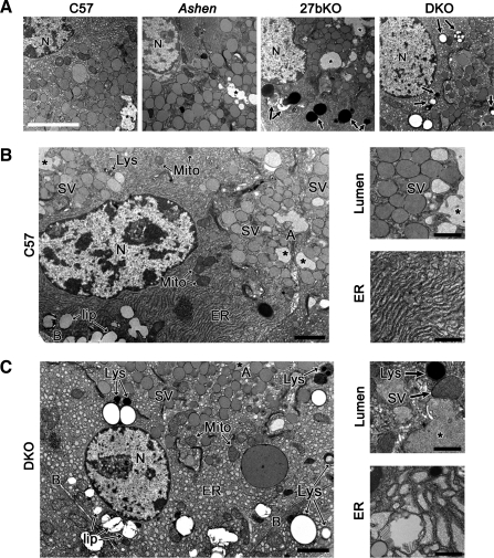
27bKO and DKO mouse LG are morphologically distinguishable from those of the C57 background strain. A: electron micrographs of 27bKO and DKO mouse LG showed major morphological differences relative to LG from the C57 background strain. Ashen LG, however, were morphologically akin to those from C57. Notable changes in 27bKO and DKO mouse LG included some loss of polarity and increased expression of degradative organelles (arrows). N, nucleus; *, lumen. Bar = 10 μm. B: LG from C57 mice showed clearly polarized acinar cells with large pools of SV located just beneath the APM (A) delineating the lumen (*). Toward the basolateral membrane (B) lies the nucleus (N), mitochondria (Mito), and lipid droplets (lip). Throughout the cytoplasm, several lysosomes (Lys) and well organized endoplasmic reticulum (ER) can be detected. High magnification images show representations of the dense SV population that lies beneath the APM and ER. C: DKO LG tissue exhibited decreased cellular organization and polarity, which varied in severity from region to region. Individual SV were less apically concentrated, whereas increased lysosomes and other apparent degradative organelles were prominent. Cells also displayed swollen or vesiculated ER as shown at higher magnification along with an example of the less densely populated subapical region beside the lumen. 27bKO morphology was similar to DKO and is not shown here. At lower magnification, bar is 2 μm; at higher magnification, bar is 1 μm.

