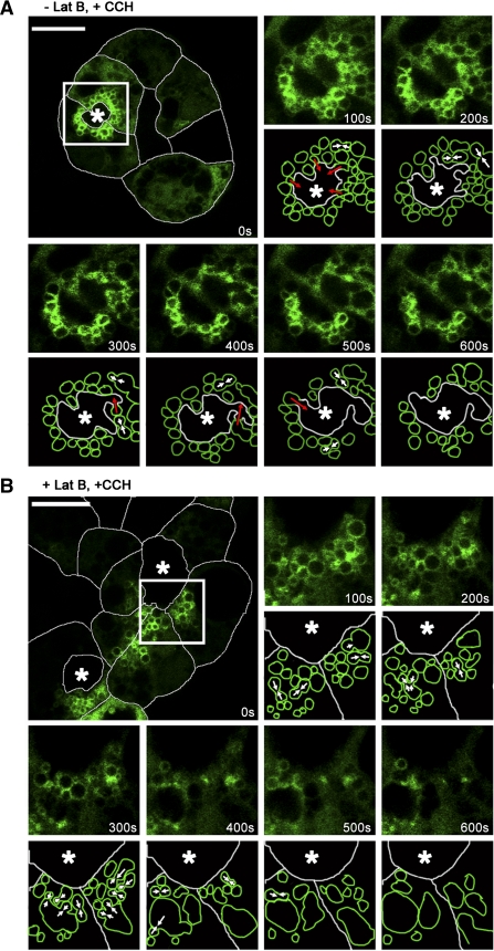
Fig. 8.
Disruption of the actin network alters terminal apical membrane fusion of Rab27b-enriched SV. A: an untreated acinus expressing YFP-tagged Rab27b was imaged in time-series immediately upon and after stimulation. Transduced cells expressing Rab27b (green) are outlined with white to delineate the cell borders. The remaining images in the series are higher magnifications of the boxed region, along with matching schematics that represent the events described. Within 100 s after CCH addition, the region around the lumen began to constrict and SV moved toward the lumen, fusing with each other (white arrows) and with the APM (red arrows). Within 600 s, ∼50% of the SV were in apparent contiguity with the lumen, suggesting discharge of contents. B: an acinus treated with 10 μM latrunculin B for 60 min at 37°C was similarly imaged. With CCH addition, SV failed to move toward the lumen. Although many SV underwent homotypic fusion (white arrows), membrane of internal fused SV failed to fuse with the APM. *, Lumen. Bar = 10 μm.

