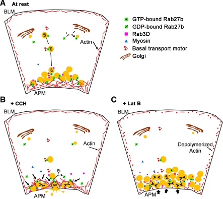
Fig. 9.
Schematic of Rab27b-enriched SV trafficking in the LG acinar cell. A: active Rab27b enriched on SV are transported to the subapical cytoplasm where they accumulate in a subapical pool as mature SV, some of which are also enriched in Rab3D. B: upon stimulation to secrete, Rab27b-enriched SV interact with actin filaments through an associated myosin motor, possibly myosin 5C. Since actin remodeling occurs as a result of stimulation to secrete, SVs are compressed with each other and undergo compound fusion, concurrent with a loss of Rab3D and release of Rab27b upon conversion of bound GTP to GDP. Simultaneously, actin compression toward the APM enables SV docking and fusion with the APM, followed by release of vesicle content into the lumen. C: when actin polymerization is disrupted, trafficking from the basal region to the subapical membrane appears intact. Upon stimulation to secrete, SV appear to accumulate beneath the APM and come into close contact with each other, which allows for some compound fusion (small arrowheads). More significantly, SV do not appear to be able to undergo fusion with the APM and the lumen shape does not change. Bold arrows represent blocked steps of the trafficking pathway.

