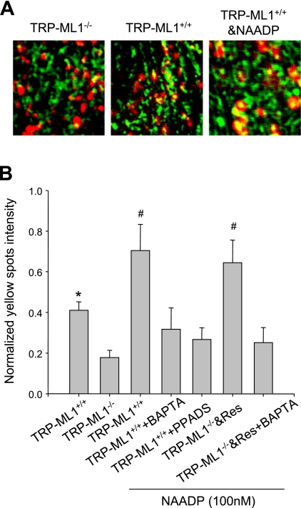
Fig. 6.
NAADP/TRP-ML1-dependent interaction of endosomes (Endo) and Lyso in human fibroblast cells by confocal microscopic assay. A: dextran-conjugated rhodamine (Rho) red shows the trace of Lyso, Oregon green 488 indicates the Endo, and the yellow spots demonstrate the mixed content between Endo and Lyso. In TRP-ML1−/− group, the relative positions of Endo and Lyso have no obvious change during the 10-min recording; consistently, there are few yellow spots observed. However, in TRP-ML1+/+ panel, the interaction of Endo and Lyso is significantly increased, and the yellow spot intensity was subsequently enhanced. When TRP-ML1+/+ is treated with NAADP, the yellow spot intensity was further elevated. B: the summary of normalized yellow spot density among different groups in 10-min image recording, which indicates that the presence of TRP-ML1 could increase the Endo and Lyso interaction and result in a significant increase of yellow intensity compared with that of TRP-ML1 deficiency cells, and NAADP could further enhance the process. Res, rescue. Values are means ± SE. *P < 0.05 vs. TRP-ML1−/− group. #P < 0.05 vs. TRP-ML1+/+-only group (n = 5∼6).

