
Figure 1.
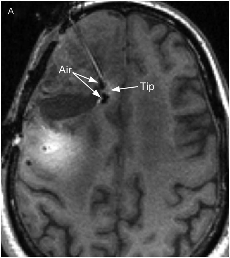
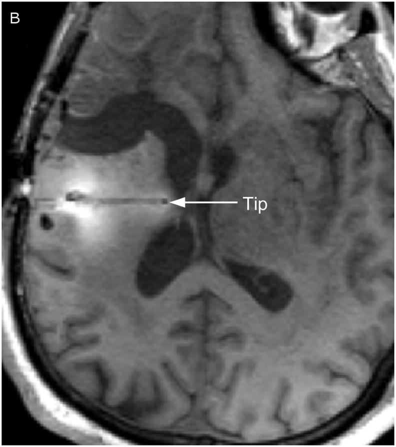
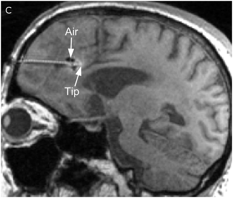
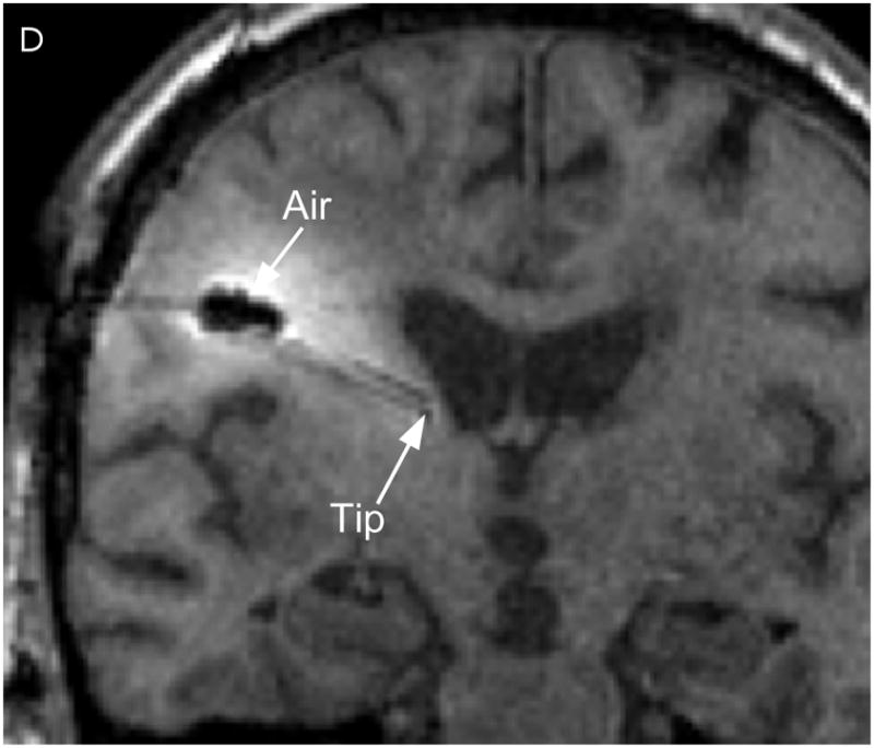
Catheter locations. A T1-weighted MRI acquisition after 24 hours of infusion was re-sliced obliquely to show the full catheter extent in two perpendicular planes. The anterior catheter is shown in two views on the left panels (A & C), and the posterior catheter in two planes on the right panels (B & D).