Fig. 1.
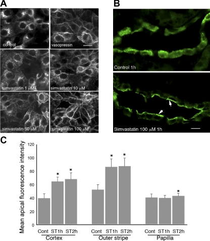
Simvastatin-induced membrane accumulation of aquaporin-2 (AQP2) in AQP2-expressing LLCPK1 cells (LLC-AQP2) and in principal cells of the collecting duct (CD) from kidney slices in vitro. A: LLC-AQP2 cells treated with ethanol (EtOH, control), vasopressin (VP, 10−8 M) for 30 min, and with simvastatin at various concentrations for 1 h. Membrane accumulation of AQP2 is seen in cells treated with VP and with simvastatin in a dose-dependent manner. Specifically, AQP2 membrane accumulation is detectable at 10 μM of simvastatin and peaks at 100 μM of simvastatin. Simvastatin-induced membrane accumulation of AQP2 was further investigated in Brattleboro rat kidney slices in vitro as shown in B. Similar to results in cell culture, treatment with simvastatin (100 μM) for 1 h causes significant membrane accumulation of AQP2 in principal cells of the CD in kidney slices (indicated by arrows), whereas in the control unstimulated state, AQP2 distributes throughout the cytoplasm. C: quantification of apical accumulation of AQP2 in kidney slices treated with simvastatin (ST). A strong and significant apical accumulation of AQP2 is seen in CD located in the cortex and outer medulla, whereas a weaker apical staining is seen in the inner medulla. Cont, control. Error bars represent means ± SE. *P < 0.01. Data were obtained from 3 experiments. Bar = 10 μm in A and B.

