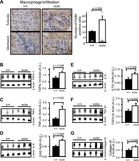
Fig. 1.
Indications of renal inflammation and signs of subtle renal injury in a mouse model of reduced β-epithelial Na channel (ENaC). A, left: representative images of renal cortical sections stained with the F4/80 antibody to identify macrophage infiltration (arrows). The sections were counterstained with hematoxylin-and-eosin. Right: group data showing that kidneys from mutant mice (m/m, n = 6) contain 2-fold more F4/80-positive cells than wild-type (+/+, n = 8). Shown is Western blotting detection of leukocyte marker target of an antiproliferative antibody (TAPA-1; B) and inflammatory cytokines TNF-α (C), IL-6 (D), IL-1β (E), as well as transforming growth factor (TGF)-β1 (F), a growth factor associated with proliferation and expansion of matrix, and collagen III (G), a marker of extracellular matrix. Protein samples were obtained from whole kidney lysates in wild-type (+/+, n = 3) and mutant mice (m/m, n = 3). β-Actin loading control is shown below the corresponding blot. Quantitative data are shown on the right. Values are means ± SE. P values for analysis with t-test are provided. *Significantly different from control, P < 0.05.

