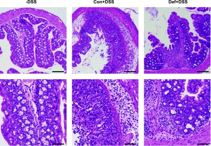
Fig. 5.
Histological images confirm human DAI scores. Hematoxylin and eosin-stained sections confirm that non-DSS animals have normal histological architecture and cytology and are not inflamed (Con and Def without DSS were identical and both are represented by the -DSS image); Con+3% had severe and diffuse destruction of the epithelial layer, neutrophil infiltration in both epithelium and lamina propria along with edema, affecting most of the tissue. Def+3% had focal inflammation in both the epithelium and lamina propria, but the tissue damage was much less severe. Scale bar = 100 μM.

