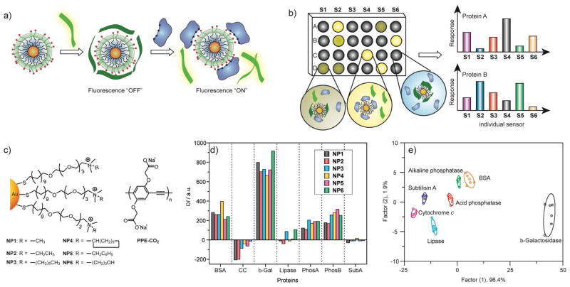
Figure 11.
a) Displacement of quenched fluorescent polymer by protein analyte with concomitant restoration of fluorescence. b) Pattern generation through differential release of fluorescent polymers from gold nanoparticles. c) Chemical structure of cationic gold nanoparticles (NP1-NP6) and anionic fluorescent polymer PPE-CO2 (n ~ 12). d) Fluorescence response (ΔI) patterns of the NP-PPE sensor array (NP1 – NP6) against various proteins (CC: cytochrome c, β-Gal: β-galactosidase, PhosA: acid phosphatase, PhosB: alkaline phosphatase, SubA: subtilisin. e) Canonical score plot for the first two factors of simplified fluorescence response patterns obtained with NP-PPE assembly arrays against 5 μM proteins. The canonical scores were calculated by LDA for the identification of seven proteins. The 95% confidence ellipses for the individual proteins are also shown.

