
Figure 12.
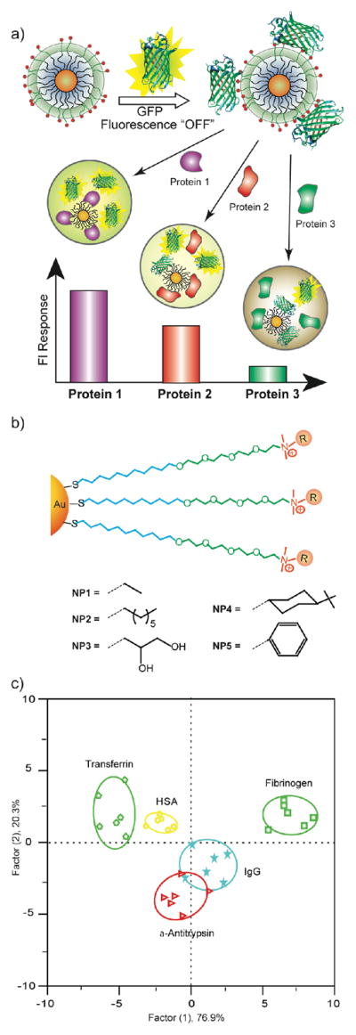
a) Schematic illustration of the competitive binding between protein and quenched nanoparticle-GFP complexes and protein aggregation leading to the fluorescence light-up or further quenching. b) Chemical structure of cationic gold nanoparticles. c) Canonical score plot for the fluorescence patterns as obtained from LDA against five protein analytes at fixed concentration (500 nM) with 95% confidence ellipses.