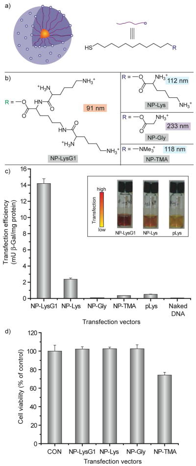
Figure 9.
a) Schematic illustration of the monolayer protected gold nanoparticles used as transfection vectors. b) Chemical structures of headgroups presented on the surface of the nanoparticles, with nanoparticle-plasmid DNA nanoplex diameters. c) Effective transfection using NP-LysG1 and NP-Lys relative to positive controls, NP-TMA and polylysine (pLys). No appreciable enzyme activity in absence of vectors. Inset showing solution color during β-Gal activity assay performed after transfection. Color change: yellow (substrate) to red (product). d) Cell viability determined by Alamar blue assay at the end of transfection showing low toxicity for amino acid-terminated ligands.

