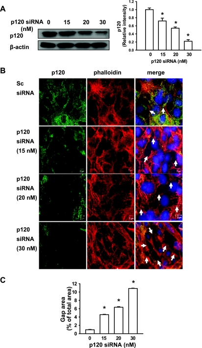
Fig. 4.
p120 siRNA increased cyclic stretch-induced gap formation in a dose-dependent manner. MLE-12 epithelial cells were transfected with scrambled and p120 siRNAs. At 48 h posttransfection, cells were then exposed to cyclic stretch (20%) for 2 h. A: the p120 siRNA specifically knocked down p120 expression as demonstrated at the protein levels in a dose-dependent manner. Left: representative Western blots of p120 protein expression. Right: the density of proteins in the control group was used as a standard (1 arbitrary unit) to compare relative densities in the other groups. *P < 0.05, compared with control groups. Data are representative of 3 independent experiments. B: after fixation and permeabilization, cells were incubated with anti-p120-catenin primary antibody followed by Alexa-conjugated secondary antibody (green). The F-actin and nucleus were stained with Alexa 546 phalloidin (red) and DAPI (blue), respectively. Scale bars = 5 μm. C: quantitative analysis of cyclic stretch-induced gap formation in alveolar epithelial cells transfected with p120 siRNA was performed as described in materials and methods. Shown are representative results of 5 independent experiments. *P < 0.05 vs. control group (static).

